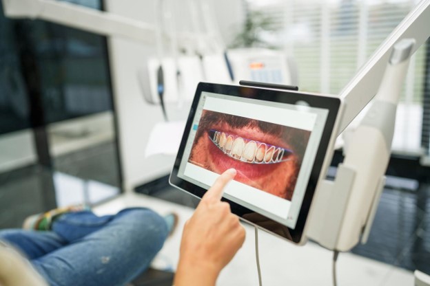
A treatment plan rendered in three dimensions looks authoritative. The implant sits in colored bone at a stated angle and a stated depth, the nerve canal is traced in a bright line, and the future restoration is already drawn on top of it. Nothing on that screen looks uncertain. Digital Implant Dentistry earns much of its reputation from exactly that clarity, and the clarity is largely deserved. It is also the reason the honest version of the subject tends to get skipped. Every element of the plan rests on data captured from a living patient in a matter of seconds, and captured data has limits. Bone can be thinner than the image suggests. Tissue compresses. Two files can merge slightly out of register. None of this makes treatment unsafe, because Digital Implant Dentistry includes a sequence of verification steps built specifically to test the plan before the plan reaches bone. What follows describes where the errors originate and what catches them.
Why a Confident Digital Implant Dentistry Plan Is Not the Same as a Correct One
Software renders every plan with the same smooth surfaces and the same crisp edges, whether the underlying scan was excellent or marginal. A volume degraded by artifact and a volume acquired under ideal conditions both produce a planning screen that looks finished. The display carries no visual warning that says the cortical boundary in one region is an interpolation rather than a measurement. This is the central caution in Digital Implant Dentistry: rendering quality and data quality are separate things, and the first one is always high. Precision and accuracy are also separate. A system can place a fixture repeatably at the coordinates it was given, which is precision, while those coordinates sit a small distance from where the anatomy actually is, which is an accuracy problem no amount of repeatability corrects. A clinician reading a plan well is therefore reading it skeptically, asking what each boundary is based on and which regions of the scan deserve less confidence than the rendering implies.
Metal Restorations, Scatter, and Artifact in Digital Implant Dentistry Imaging
Existing crowns, posts, amalgam restorations, and orthodontic retainers all interact with the beam in ways that corrupt the reconstruction around them. Dense metal absorbs low energy photons preferentially, which shifts the measured density of everything behind it, and the reconstruction resolves that inconsistency as dark bands, bright streaks, and cupping across the surrounding region. The damage is local but not trivial, because the region next to a heavily restored tooth is frequently the exact region where an implant is planned. Artifact in Digital Implant Dentistry can make a thin buccal plate look absent, make a healthy trabecular pattern look sclerotic, or hide a small radiolucency entirely. Adjusting head position, narrowing the field of view, and selecting acquisition settings suited to a restored mouth all reduce the effect, and correction algorithms help, but none of these eliminates it. The practical safeguard is recognition. A clinician who identifies a streak as an artifact treats that area as unmeasured rather than as measured and reassuring.
Patient Movement During a Scan and What It Does to Digital Implant Dentistry Data
A cone beam acquisition takes several seconds, during which the source and detector rotate around the head and record a series of projections. The reconstruction assumes the anatomy held perfectly still for all of them. A swallow, a deep breath, a slight settling of the shoulders, or an involuntary tremor breaks that assumption. The result is not usually an obvious blur. It shows up as doubled cortical outlines, softened edges where a boundary should be sharp, or subtle stair-stepping through a series of slices, and a measurement taken from a softened edge inherits the softening. Motion is one of the quieter error sources in Digital Implant Dentistry because small movements produce images that still look acceptable at a glance. Chin rests, head straps, bite blocks, clear coaching before the exposure, and a short practice run all reduce the risk. When a reviewer sees the signs anyway, the correct response is a repeat acquisition rather than careful measurement of unreliable data.
What a Cone Beam Scan Does Not Resolve in Digital Implant Dentistry
Every volume is built from voxels, and no structure smaller than the voxel can be represented honestly. A buccal plate thinner than the sampling resolution may vanish from the image, reading as a dehiscence that does not exist, or it may be smeared across neighboring voxels and read as thicker bone than is really present. Partial volume averaging is responsible for both outcomes. Fine neural anatomy presents a parallel limitation in Digital Implant Dentistry, since the inferior alveolar canal is traceable where its walls are well corticated and becomes ambiguous where they are not. Accessory canals, incisive extensions, and the anterior loop of the mental nerve are variable in size and sometimes sit below what the scan can show. Increasing resolution increases radiation dose, so the choice of settings is always a compromise. The workable response is to plan with deliberate clearance from structures whose margins the image describes approximately rather than exactly.
Soft Tissue Thickness and the Blind Spot in Digital Implant Dentistry Imaging
Cone beam imaging is configured for hard tissue, and that configuration comes at the cost of soft tissue detail. Mucosa, attached gingiva, and the lip and cheek tend to blend together at similar density values, and the tissues are often displaced by whatever holds the mouth in position during the exposure. The consequence is a plan that treats the bone crest as the reference point while the restoration will actually emerge through a tissue layer of unknown thickness. Depth decisions in Digital Implant Dentistry depend on that layer, because a platform placed at an ideal depth relative to bone can sit too shallow or too deep relative to the eventual gingival margin. Direct measurement solves what the scan does not, whether by sounding the tissue with a periodontal probe under anesthetic, by acquiring a scan with the tissues retracted, or by using a radiographic appliance that separates the lip and cheek from the ridge during acquisition.
Merging Surface and Volumetric Data in Digital Implant Dentistry
Two very different datasets have to be combined before planning can begin. The volumetric scan supplies bone and internal anatomy at moderate resolution. The optical surface scan supplies crowns, gingival contours, and occlusal detail at far finer resolution. Registration aligns them by matching shared landmarks, usually cusp tips, incisal edges, and other stable tooth surfaces that appear in both. The alignment can drift when few natural teeth remain, when the available landmarks are clustered on one side, or when the same metal artifact that degrades the volume also degrades the surfaces used to register it. A merge in Digital Implant Dentistry fails quietly rather than loudly, since a mismatched superimposition still renders as one clean model. Verification means checking the overlay in axial, sagittal, and coronal slices, and specifically on teeth that were not used as registration landmarks, to confirm that the surface outline tracks the volumetric outline everywhere and not just where the software was told to match.
Edentulous Arches, Mobile Tissue, and the Limits of Digital Implant Dentistry Scanning
An edentulous arch removes nearly everything a scanner relies on. There are no cusps, no embrasures, and no fixed landmarks to anchor the stitching process, only a broad ridge of similar looking mucosa. Optical scanners assemble a model by matching overlapping frames, and a featureless surface gives them little to match, so small errors accumulate across the arch and distort the overall dimension in ways that are hard to detect from the model itself. The tissue compounds the problem, because mucosa is mobile and displaceable. It moves under the scanner tip, under retraction, and under the pressure of a prosthesis, meaning the recorded surface depends partly on how it was recorded. Digital Implant Dentistry handles this with references that do not move, including fiducial markers on a stable appliance, a well fitting existing denture used as a scan reference, and bone supported rather than tissue supported guide designs where the anatomy allows for them.
When a Guide Fits the Model but Not the Mouth in Digital Implant Dentistry
A surgical guide designed against a digital model will seat on that model flawlessly, and it will seat on a printed version of that model just as well. The mouth is a different object. Printing introduces dimensional error through resin shrinkage, layer thickness, build orientation, incomplete post-curing, and the finishing required to remove supports. Sterilization can add a small amount of distortion of its own. Tissue supported designs then rest on mucosa that compresses unevenly under hand pressure, so the same guide can settle to slightly different depths depending on how it is held. The consequence for Digital Implant Dentistry is that a guide which rocks even slightly on a fulcrum is no longer reporting the position it was designed to report, and the deviation grows along the length of the osteotomy. Undercuts, residual interproximal material, and inadequate relief in the design are all common reasons a technically correct guide seats imperfectly on a real arch.
The Tolerance Stack in Digital Implant Dentistry
Each individual source of error discussed so far is small, and each one alone would be clinically unimportant. The difficulty is that they do not occur one at a time. Voxel resolution, a trace of patient motion, registration drift, fabrication tolerance, the clearance between sleeve and drill, seating variability, drill flex, and deflection off a dense cortical layer all contribute in the same procedure. They can partially cancel, which is fortunate, or they can align in the same direction, which is not. Angular error compounds the arithmetic further, since a deviation that is negligible at the platform becomes meaningfully larger at the apex of a long fixture. The reasoning that protects a case in Digital Implant Dentistry is therefore cumulative rather than item by item. Safety margins to the nerve, the sinus, and adjacent roots are set against the worst plausible combination of tolerances, not against the number shown on screen, which represents the plan rather than the outcome.
Verifying Guide Seating Before the First Drill in Digital Implant Dentistry
The most important verification step costs almost nothing and happens before any bone is touched. The guide is tried in and evaluated deliberately. Inspection windows designed into the body of the guide let the clinician confirm visually that tooth surfaces are fully contacting the intended seating areas with no gap and no trapped tissue. Alternating finger pressure at several points tests for rocking, and the guide should not pivot or spring back anywhere. Repeatability matters as much as fit, so the guide is removed and reseated to confirm it arrives in the same position each time. Anchor pins add fixation where the seating surface is limited. Digital Implant Dentistry treats a failed seating check as decisive information rather than as an inconvenience, because a guide that does not seat identically twice cannot transfer a plan accurately. The appropriate responses are to correct the interference, to proceed with the guide as a reference only, or to reschedule.
Open Flap Access as a Check on Digital Implant Dentistry Assumptions
Raising a flap converts an inference into an observation. Once the ridge is exposed, its actual width, the actual crest height, and the presence of a buccal concavity or a knife edge profile are all directly visible and directly measurable with a caliper. That is a fundamentally different category of information than a boundary interpreted from a grayscale image, and it is the most reliable available check on what the scan implied. Flapless approaches have genuine advantages in comfort, bleeding, and healing time, and they remain appropriate when the imaging is clean and the anatomy is generous. Digital Implant Dentistry becomes more conservative as the imaging becomes less certain, and a region obscured by artifact, a plate that may or may not exist at the resolution used, or a ridge with a suspected undercut are all reasonable grounds to open and look. Exposure also permits immediate grafting if the exposed anatomy turns out to differ from the plan.
Torque and Tactile Feedback as Information No Digital Implant Dentistry File Contains
Osteotomy preparation generates a continuous stream of information that exists nowhere in any dataset. Resistance changes as the drill passes from dense cortical bone into softer marrow, and an experienced operator reads those transitions through the handpiece. A sudden loss of resistance indicates that the drill has entered a space it should not have entered, whether a large marrow void, a sinus, or a perforation through a plate, and it is felt at once rather than discovered later. Insertion torque then reports whether the fixture achieved primary stability in that particular bone, and low torque can reveal that the osteotomy was slightly oversized for the density actually encountered. Radiographic density estimates bone quality but does not measure it, so Digital Implant Dentistry depends on this tactile layer as an independent confirmation from a source that cannot inherit any imaging error. When the feel of the bone disagrees with the plan, the feel of the bone is the more current evidence.
Intraoperative and Post-Placement Radiographs as Confirmation in Digital Implant Dentistry
Imaging returns as a checkpoint during the procedure rather than only before it. A periapical radiograph taken with a depth gauge or a direction indicator seated in a partially prepared osteotomy shows trajectory relative to adjacent roots and to the canal while the path can still be adjusted, which is the whole point of taking it early. A film after placement confirms the final depth, the angulation, and the complete seating of the healing component, and it establishes the baseline against which future bone levels will be compared. Comparison with the planned position documents any deviation for the record. These images verify rather than certify, because a two dimensional radiograph cannot report buccal or lingual position, so Digital Implant Dentistry treats them as one layer among several rather than as a final word. A post-placement volumetric scan supplies three dimensional confirmation when a specific question genuinely warrants the additional exposure.
Abandoning the Plan Mid-Procedure as Good Digital Implant Dentistry Practice
Every plan is a hypothesis about anatomy that has not yet been seen directly. When the operative findings contradict it, the correct action is to follow the findings. That can mean selecting a narrower or shorter fixture than planned, shifting position within the available bone, grafting the site and returning after healing, or closing and rescheduling with better imaging and a revised design. None of these outcomes is common, and none is a defect in the technology. Patients occasionally interpret a change of course as evidence that something went wrong, when in practice it is the moment the safeguards did their job. The verification steps that make Digital Implant Dentistry dependable have value only if the clinician is genuinely prepared to act on what they reveal, including at an inconvenient point in the appointment. A practice that discusses this possibility during consultation, before anything is scheduled, is describing normal contingency planning rather than warning of trouble.
Why Patients Choose August de Oliveira DDS for Digital Implant Dentistry
Dr. August de Oliveira has built his practice around digital dentistry, and the Encino office runs both chairside CEREC workflows and full laboratory workflows depending on what a given case requires. Working in both modes regularly gives the office direct familiarity with where scan data holds up and where it needs to be checked against the mouth, which is precisely the judgment this subject depends on. Digital Implant Dentistry at this practice is presented to patients with its verification steps included rather than as a purely automated result, and questions about imaging quality, guide fit, and contingency options are treated as reasonable things to ask before treatment begins. The office is located at 5400 Balboa Blvd #231, Encino, CA 91316, and sees patients Monday through Thursday from 8 to 5, with Fridays by appointment. Patients travel to the practice from Encino, Sherman Oaks, Van Nuys, Tarzana, Calabasas, Woodland Hills, Studio City, and Reseda.
Final Thoughts on Digital Implant Dentistry
The useful conclusion is not that digital planning is unreliable. It is that the plan and the patient are two different things, and the entire structure of modern implant surgery exists to keep the distance between them small. Scans have resolution limits, tissue moves, files merge imperfectly, and guides made from resin are physical objects with physical tolerances. Against all of that sit the seating check before the first drill, direct visualization when the imaging leaves a question, the feel of the bone during preparation, torque at insertion, and radiographic confirmation during and after placement. Digital Implant Dentistry is dependable because those checks are performed, not because the software is assumed to be right. This article is general information and is not a treatment recommendation for any individual case. Patients in Encino and the surrounding San Fernando Valley who want implant treatment planned carefully and verified at every stage can call August de Oliveira DDS at 5400 Balboa Blvd #231, Encino, CA 91316 to schedule a consultation.