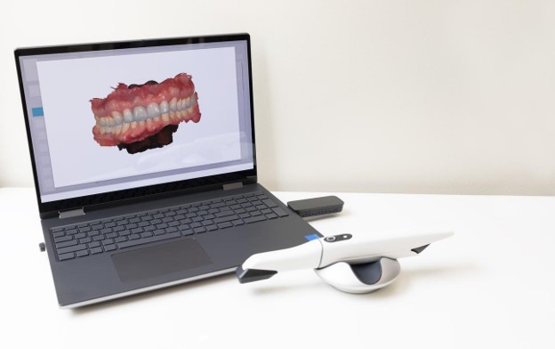
Most conversations about digital implant dentistry stop at the day of surgery. The scan is taken, the plan is built, the guide is printed, the implant goes in, and the story ends. The more useful story starts later, in year five or year twelve, when a crown fractures on a Friday afternoon, or when a clinician who never met the patient must order a component for a fixture placed by someone who has since retired. What determines how easy or how miserable that day becomes is not the surgery. It is whether a readable, complete, organized digital file still exists. Treatment done with digital implant dentistry produces an archive as a byproduct, and that archive quietly carries most of its value into the future. August de Oliveira DDS in Encino treats those files as part of the restoration itself, because digital implant dentistry is only finished when the record is stored in a form someone can actually open and use.
What Digital Implant Dentistry Actually Produces as a Permanent Record
A completed case generates a specific and surprisingly durable set of artifacts. There is the cone beam volume taken before treatment, usually stored as DICOM. There are intraoral scans of the arch before preparation and after delivery, typically exported as STL or PLY. There is the planned implant position expressed in software coordinates relative to the surrounding anatomy. There is the surgical guide design, the abutment design, and the final prosthesis design, each a mesh file that can be reopened and re-milled, plus the shade record and the photographic series. There is the hard data on the fixture itself, meaning system, diameter, length, and connection platform. Each of these becomes a file under digital implant dentistry rather than a memory or a handwritten note in a chart that may not survive a practice sale. Taken together they describe not just what was done but how to reproduce it, which is a different and more valuable thing.
CBCT Volumes and Pre-Treatment Scans as the Anatomical Baseline of Digital Implant Dentistry
The pre-treatment cone beam volume is a three dimensional snapshot of bone, sinus floor, nerve canal, and adjacent root positions on a specific date. Years later that volume answers questions no new image can answer, because a new image shows only the present. Comparing the original volume to a current one reveals whether a sinus graft consolidated, whether a buccal plate thinned, or whether an adjacent tooth developed a new lesion. The pre-treatment intraoral scan does something parallel for the soft tissue and the occlusion, recording gingival contours and the position of every opposing cusp before anything changed. Both are stored by digital implant dentistry as neutral, standards based files rather than as screenshots inside a planning application. A DICOM series opens in essentially any modern imaging viewer, and an STL of the arch imports into any design software. That portability is what makes the baseline useful to a clinician who is not the one who created it.
The Planned Position and Surgical Guide Design Stored by Digital Implant Dentistry
The treatment plan file holds the implant position as it was intended, described in relation to the ridge, the opposing arch, and the planned restoration. The guide design holds the physical expression of that plan, including sleeve position, depth stops, and the surfaces the guide seated against. Both remain useful long after the guide has been thrown away. If a second implant is later planned in the same quadrant, the original plan file shows the angulation and depth logic already in place. If a guide needs to be reprinted because a staged case resumed after a delay, the design is simply reopened and sent to the printer again. A case handled with digital implant dentistry also allows the planned position to be compared against a post-placement radiograph or scan, which documents the actual as-placed result. That comparison matters when someone in the future needs to know whether the fixture sits where the prosthetic design assumes it sits.
Implant System, Diameter, Length, and Platform in the Digital Implant Dentistry Record
The single most consequential line in the whole record is the one naming the fixture. System, diameter, length, connection type, and platform diameter together determine which healing abutments, impression components, scan bodies, titanium bases, screws, and drivers will fit. Get any of those wrong and a component arrives that seats imperfectly or not at all. Those specifications are tied by digital implant dentistry to the case file and to the design files that depend on them, so a titanium base ordered eight years later matches the connection the crown was designed around. The same information supports component ordering directly, since manufacturers and distributors need the exact reference to ship the right part, and it supports any warranty or replacement program the manufacturer operates, which almost always requires proof of what was placed and when. A record without the fixture specifications is a record that still leaves the next clinician guessing.
Shade and Photographic Documentation Within Digital Implant Dentistry
Shade is easy to underestimate until a central incisor crown has to be remade next to a natural tooth that has aged for a decade. A recorded shade, captured with calibrated photography alongside a reference tab, tells the ceramist what the original match was built from. The photographic series adds surface texture, translucency at the incisal edge, characterization, and the position of the gingival zenith. That visual layer is worth a great deal to digital implant dentistry, because a mesh file describes shape but says nothing about color or optical behavior. Photographs also document the emergence profile and the shape of the tissue collar that was developed during provisionalization, which is information no scan of the finished crown fully preserves. When a replacement is needed years later, the difference between a crown that disappears and a crown that reads as a restoration often comes down to whether that photographic record was captured and kept.
Remaking a Lost or Broken Crown From a Stored File in Digital Implant Dentistry
Consider the ordinary emergency. A patient bites into something hard and the implant crown fractures. Without a record, the appointment involves removing the remaining restoration, identifying the connection, finding a matching scan body, rescanning the arch, redesigning the crown, and either milling it or sending it out. With a stored design file, the crown is already designed. The file is reopened, verified against a current scan for any drift in the neighboring teeth or the opposing arch, and sent to the mill. Here digital implant dentistry compresses a multi-appointment problem into something that can often be handled in a single sitting, because the intellectual work of the original case was saved rather than discarded. The saved design also means the replacement reproduces the contacts, contours, and occlusal scheme the patient has been functioning with, instead of introducing a new shape that has to be adapted to all over again.
How Digital Implant Dentistry Preserves the Original Tooth Shape for Later Replacement
There is a specific and underappreciated use for the scan taken before the natural tooth was removed. That file contains the patient’s own anatomy, meaning the exact width, height, incisal edge position, embrasure form, and axial contours of the tooth that was lost. A replacement designed from that scan reproduces something rather than approximating it. Without it, even a skilled technician is reconstructing from adjacent teeth, mirrored contralateral anatomy, and judgment. In digital implant dentistry that pre-extraction scan is cheap to capture and permanently reusable, and it is the reason a crown delivered a decade after the extraction can still look like the tooth that was there. The same file supports later revisions too. If a crown is remade after gingival recession changes the emergence, the original contour data gives the designer a reference for what the patient looked like before anything moved.
The Unidentified Implant Problem That Digital Implant Dentistry Prevents
One of the most expensive and frustrating situations in restorative practice involves a patient with an implant nobody can identify. The fixture was placed fifteen years ago and the surgeon has retired, sold the practice, or moved out of state. The records were paper and were purged, or they were digital and were lost in a software migration. The patient remembers nothing beyond the year. Now an abutment screw has loosened or a crown has failed, and no one knows which driver, which screw, or which restorative component will fit. Practiced with the archive in mind, digital implant dentistry makes that scenario simply not happen, because the system and specifications were recorded at the time and stored with the case. The cost of recording that information at placement is a few seconds. The cost of not recording it can be an entire diagnostic detour, and occasionally the removal of a perfectly healthy, perfectly integrated fixture.
How an Unknown Fixture Gets Identified Without Digital Implant Dentistry Records
When the record is missing, identification becomes detective work. It begins with a well angled periapical radiograph taken perpendicular to the long axis of the fixture, because the diagnostic features are geometric. Clinicians compare thread pitch and thread profile, the shape of the apex, the presence or absence of an apical chamber, the collar design, and above all the internal connection silhouette, meaning whether the platform reads as an external hex, an internal hex, a tri-channel, a conical connection, or something else. Software identification tools and published reference atlases match those features against known systems, and trial fitting of candidate components sometimes settles it. The process often succeeds, but it consumes appointment time, may require ordering several components on speculation, and occasionally ends inconclusively. The entire exercise is avoided by digital implant dentistry, which writes down at placement what would otherwise be reverse engineered years later from a shadow on a radiograph.
Baseline Radiographs and Bone Level Comparison in Digital Implant Dentistry
Peri-implant bone loss is only measurable against a starting point. A radiograph taken at delivery, with a reproducible angle and a known reference dimension such as thread pitch or platform width, establishes where the bone crest sat when function began. Every subsequent radiograph is then interpretable. Two millimeters of bone below the platform means one thing if the bone was at the platform at delivery and something else entirely if it was already two millimeters apical at the start. This is supported in digital implant dentistry by pairing the delivery radiograph with a delivery scan, which captures soft tissue height and contour at the same moment. Without a baseline, a clinician meeting the patient years later can describe the current state but cannot describe the trend, and the trend is what actually drives decisions about whether to monitor, intervene, or restore differently.
Full Arch Prostheses, Speech, and Bite Adaptation in Digital Implant Dentistry
Full arch cases raise the stakes considerably. A patient who has worn a fixed full arch prosthesis for years has adapted their speech, tongue posture, lip support, and chewing pattern to that exact shape. Tooth position, palatal contour, flange thickness, and vertical dimension are all things the neuromuscular system learned. When such a prosthesis fractures or wears through, remaking it from scratch means the patient relearns everything, and some patients never fully adjust. A stored design file removes that problem. The same geometry is milled again in a fresh material, and the replacement feels like the one that broke. Working from that saved baseline, digital implant dentistry also allows deliberate, incremental modification, so if the patient wanted shorter incisal edges or a small change in phonetics, the design is edited rather than rebuilt. The reference shape is preserved either way, which is the point.
Transferring Digital Implant Dentistry Records Between Practices
Patients move, change insurance networks, and change dentists. When they do, the question is whether the record moves with them. Files stored in open, standards based formats travel easily. A DICOM volume, an STL or PLY of each scan, a PDF or image of the implant specifications, and the photographic series can be copied to secure storage and handed to any receiving office. Files locked inside a proprietary planning or design environment sometimes cannot, or can only be exported in a degraded form. The record built by digital implant dentistry is most valuable when the patient is treated as the owner of the data. Anyone leaving a practice should ask for the pre-treatment and post-delivery scans, the cone beam data, the final restoration design file, the implant brand and specifications, and the delivery radiograph. Receiving those in readable formats means the next clinician starts from the actual history rather than from a fresh set of assumptions.
Revision Planning, Add-On Implants, and Provisional Duplication in Digital Implant Dentistry
Cases grow. A patient restored with two implants may need a third when a neighboring tooth fails. Planning that new fixture against the as-placed positions of the existing ones, rather than against a guess, controls angulation, parallelism, and prosthetic space from the beginning. A stored post-placement scan with scan bodies, or a post-placement cone beam volume, gives that as-placed geometry. Provisionals are also preserved by digital implant dentistry, which matters more than it sounds. When a patient has worn and approved a provisional shape, that approved geometry is a specification. Copying it forward into the definitive design means the final restoration delivers what the patient already approved rather than a new interpretation. The same duplication logic protects against provisional failure, since a fractured temporary can be reprinted or remilled from the saved file rather than rebuilt chairside from scratch.
The Honest Limits of Digital Implant Dentistry Archives and What to Ask a Practice
None of this works automatically. A file only helps if it was actually saved, actually backed up in more than one location, and actually stored in a format that will still open years from now. Practices change software, migrate servers, and sometimes lose data in transition. Proprietary formats that only one application can read are a genuine risk, which is why open standards like DICOM for imaging and STL for meshes matter so much. On its own, digital implant dentistry does not guarantee retention, and retention policies vary by office and by law. Patients are entitled to ask direct questions. What is archived, in what formats, backed up where, and can copies be released on request. It is equally reasonable to ask for the implant brand and specifications card at the end of treatment, and to keep a copy somewhere personal, such as with important documents or in a secure cloud folder.
Why Patients Choose August de Oliveira DDS for Digital Implant Dentistry
Dr. August de Oliveira has built his practice around digital dentistry, and the Encino office runs both chairside CEREC workflows and full laboratory workflows depending on what a case calls for. That combination matters for the archive, because a case designed digitally produces design files whether it is milled in the operatory or finished by a laboratory. The office keeps records in standard formats and documents implant specifications as part of the treatment record, so patients are not left holding a restoration nobody can identify later. At this practice, digital implant dentistry is treated as a long horizon commitment rather than a single procedure, with the same attention given to what happens at year ten as to what happens on placement day. The office is located at 5400 Balboa Blvd #231, Encino, CA 91316, and serves patients from Encino, Sherman Oaks, Van Nuys, Tarzana, Calabasas, Woodland Hills, Studio City, and Reseda.
Final Thoughts on Digital Implant Dentistry
The value of an implant is not settled on the day it is placed. It is settled every time something needs attention afterward, and on each of those days the deciding factor is how much of the original case still exists in usable form. A stored crown design turns an emergency into a short appointment. A pre-extraction scan lets a replacement reproduce a tooth instead of inventing one. A recorded fixture specification removes an entire category of expensive uncertainty, and a saved full arch design protects speech and function that took months to establish. Most of the reputation of digital implant dentistry is earned during planning and placement, but it repays patients quietly for decades through the file it leaves behind.
Patients in Encino and the surrounding San Fernando Valley who want implant treatment documented properly can contact August de Oliveira DDS at 5400 Balboa Blvd #231, Encino, CA 91316. The office sees patients Monday through Thursday from 8 to 5, with Fridays by appointment. Call to schedule a consultation and discuss what digital implant dentistry can preserve for the years ahead.